Table of Contents
- Introduction
- What is the Cardiac Conduction System?
- How Conduction Systems Controls Heartbeat?
- Components of the Cardiac Conduction System
- Function of the Heart Conduction System
- Steps in the Heart Conduction Pathway
- Conclusion
The heart is an extraordinary organ that continuously pumps blood throughout the body. The normal range is considered between 60 and 100 beats per minute. In case it goes down below 60, it’s called bradycardia or a slow heart. If it shoots over 100, it’s known as tachycardia or a fast heart. However, remember that a healthy heart rate can change depending on what you’re doing. So, what regulates this whole mechanism? It’s the cardiac conduction system that keeps this rhythm in check and ensures your heart beats just right for every moment.
In this blog, we will discuss the conduction system of the heart, its importance, functions, and the mechanism of the heart conduction pathway.
Master ACLS Now
Get ACLS certified with confidence
What is the Cardiac Conduction System?
The heart conduction system consists of a specialized network of cells and fibers in the heart’s wall. This system controls and manages heartbeats. It produces and transmits electrical signals that cause different parts of the heart to expand and contract and regulate the heart’s pumping action.
The contraction and expansion control the flow of blood through your heart as well as your body. There are two types of cells, namely muscle cells and conducting cells, that control the heartbeat. Apparently, conducting cells move the electric signals, while muscle cells are responsible for the heart’s contraction.
The conduction system of the heart sends signals to the brain to start a heartbeat. This system of expanding and contracting regulates the blood flow properly through the heart and body and provides the heart with an automatic and rhythmic beat.
How Conduction Systems Controls Heartbeat?
The conduction pathway of the heart helps circulate nutrient enriched blood and oxygen throughout your body. But when it fails to work properly, it affects the other organs and body parts. For example, chronic heart rhythm disorders are associated with an increased risk of dementia, with studies showing a 40-60% higher risk of developing cognitive impairments.
- The conduction pathway starts with the sinoatrial (SA) node, which acts like a tiny pacemaker in the top part of your heart and sends out an electrical signal. This signal makes the upper parts of your heart (atria) contract and push blood into the lower parts (ventricles).
- The signal then goes to the atrioventricular (AV) node, which pauses it for a moment to make sure the ventricles are full of blood.
- The signal travels through special pathways called the His-Purkinje network which makes the ventricles contract and pump blood to your whole body.
This system keeps your heart beating smoothly and in the right order, making sure blood flows properly. Heart rate is central to the process of the conduction system of the heart as the function of the heart is closely connected with the stroke volume, i.e., the amount of blood that is pumped out with each beat of heart as well as with the heart rate.
Read More: Identifying and Treating First-Degree AV Block (First-Degree Heart Block)
Components of the Cardiac Conduction System
For the cardiac conduction system to perform effectively, there are a few components that you should know. They are as follows:
-
Sinoatrial (SA) Node
The sinoatrial node or sinus node comprises a small mass of specialized tissues that are set in the upper right chamber of the heart. It generates electrical impulses to regulate the heart’s rate and rhythm.
-
Atrioventricular (AV) Node
The atrioventricular node, or AV node, is a small and compact structure in the heart that electrically connects the ventricles and the atria and allows them to beat in coordination.
-
Bundle of His
The bundle of His is a specialized cluster of muscle fibers in the cardiac conduction system diagram that carries electrical impulses between the atria and ventricles. HIS bundle is also known as the atrioventricular bundle.
-
Purkinje Fibers
Purkinje fibers are the specialized nerve cells that send electrical signals to the ventricles, making them contract. This helps the heat to maintain a consistent rhythm and coordinated contraction of its ventricles.
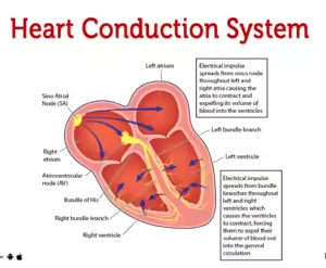
Check out this simple conduction system of heart diagram to better understand the components.
Function of the Heart Conduction System
The cardiac conduction system coordinates and regulates the rhythmic contraction of the heart muscle, ensuring efficient blood circulation throughout the body. So, after understanding what a cardiac conduction system is, let’s discuss how it works.
-
Role of Nodes, Specialized Cells, and Electrical Signals
The heart conduction pathway is composed of a network of specialized cells, nodes, and electrical signals that work together to maintain the heart’s rhythm.
- Nodes are groups of cells that can form either muscle or nerve tissue. They control electrical pulses and regulate the heartbeat. There are two types of nodes: the Sinoatrial (SA) node and the Atrioventricular (AV) node.
- The SA node is often called the heart’s pacemaker because it generates electrical signals that direct the atria to contract. The AV node is situated in the triangle of Koch, near the heart’s central area. It receives signals from the SA node and passes them to the ventricles, causing them to contract and pump blood.
- Specialized cells in the heart’s muscular wall, known as cardiomyocytes, are branched, striated muscle cells with a single nucleus found only in the heart. These cells contain contractile units that power the heart’s beating. During each beat, the cells contract together to pump blood through the heart and out to the body.
-
Heart Contractions
The electrical conduction system of the heart controls its rhythm and rate by sending electrical signals throughout the heart. The Sinoatrial (SA) node generates electrical impulses 60 to 100 times per minute. These impulses spread to the atria, causing them to contract and pump blood into the ventricles.
The Atrioventricular (AV) node slows down the spread of these impulses. The signal is then carried forward by the bundle of His, through the bundle branches, and via the Purkinje fibers, causing the ventricles to contract.
Note: Understanding how the cardiac conduction system works is essential for anyone interested in maintaining heart health. To further enhance your knowledge and be prepared for cardiac emergencies, consider enrolling in an ACLS certification course.
-
Blood Flow Regulation
The cardiovascular system regulates the velocity and amount of blood flow throughout the body in response to different stimuli. The heart contains four valves that ensure blood moves in the correct direction and prevent backflow. These valves are:
- Tricuspid Valve: Controls blood flow between the right ventricle and right atrium.
- Pulmonary Valve: This valve regulates blood flow from the right ventricle to the pulmonary arteries, which carry blood to the lungs.
- Mitral Valve: Allows oxygen-rich blood from the lungs to pass from the left atrium to the left ventricle.
- Aortic Valve: This valve opens the path for oxygen-rich blood to move from the left ventricle into the aorta, the largest artery in the body.
Each of these components plays a crucial role in maintaining the heart’s function and ensuring efficient blood circulation throughout the body.
Read more: Study Guide To Body Systems: ACLS Certification Resource
Steps in the Heart Conduction Pathway
The conduction system of the heart contains the following sequential steps of electrical events that take place during one complete contraction of the heart muscle.
-
Sinoatrial (SA) Node Excitation Signal
The Sinoatrial (SA) node is a cluster of specialized cells or pacemaker cells. It is located in the right atrium’s upper wall, which is the right upper chamber of the heart, at the junction where the superior vena cava enters.
The function of these pacemaker cells is to generate spontaneous electrical impulses that start the heartbeat. The Autonomic nervous system includes the Sympathetic and Parasympathetic nervous systems. The sympathetic nervous system makes your SA node work faster and helps to increase heart rates. The Parasympathetic nervous system makes your SA node work slower, decreasing your heart rate. This controls how fast or slowly SA nodes convey electrical signals.
-
Atrial Contraction
When your heart beats, it goes through a cycle to pump blood efficiently. First, the top chambers (atria) squeeze to push blood into the bottom chambers (ventricles). This helps fill the ventricles with blood so they can pump it out to your body. After squeezing, the atria relax.
During this squeezing phase, blood flows through valves like the mitral valve on the left side and the tricuspid valve on the right side. This equalizes the pressure between the atria and ventricles. This phase is called diastasis, and it lets the ventricles fill up properly.
Then, the atria squeeze harder, pushing more blood into the ventricles. This extra push is called atrial contraction or atrial systole. It helps your heart pump blood effectively to your lungs and the rest of your body.
Both sides of your heart left and right, work together to keep your blood flowing smoothly and keep your body healthy.
-
Atrioventricular (AV) Node
After electrical impulses spread throughout the atria, they come together at the atrioventricular node, located within the atrioventricular septum near the opening of the coronary sinus.
The AV nodes delay the impulses by approximately 120ms to ensure that the atria get enough time to eject blood completely before the ventricular systole. Then, the wave of excitation moves from the atrioventricular node into the atrioventricular bundle.
-
Bundle of His and Purkinje Fibers
The Bundle of His is a continuation of the AV node’s specialized tissue, and it transmits the electrical impulse from the AV node to the Purkinje fibers of the ventricles.
It comes down the membranous part of the interventricular septum before emerging into the two main bundles, namely the right bundle branch, which conducts the impulse to the Purkinje fibers of the right ventricle, and another is left bundle branch, which conducts the impulse to the Purkinje fibers of the left ventricle.
-
Ventricular Contraction
Ventricular contraction occurs when the two lower chambers, called ventricles of the heart, contract. This condition happens when an abnormal contraction signal called depolarisation originates from the ventricles instead of pacemaker cells.
As the ventricles contract, pressure also increases within them and exceeds the pressure in the atria, which causes the atrioventricular valves to shut.
Final Thoughts
The cardiac conduction system is a network of specialized muscle cells in the wall of the heart that sends electrical signals to the rest of the heart muscle, making it contract and relax. This system, made up of special muscle cells, helps the heart beat properly by managing its electrical signals.
For example, knowing about key parts like the SA node, AV node, and the bundle of His and Purkinje fibers helps us understand how the heart keeps a steady rhythm and pumps blood. In emergencies, this knowledge can help people recognize early signs of trouble and get medical help quickly.
Being aware of how the heart’s electrical system works can encourage healthier lifestyle choices. By keeping a heart-healthy lifestyle and learning about heart function, people can lower their risk of heart disease and improve their overall heart health.