Electrocardiography, commonly known as ECG, is a diagnostic tool recording the heart’s electrical activity. You can get valuable information about the heart’s rhythm, rate, and any abnormalities that may be present. ECG waves explained in the right manner can help healthcare professionals act fast in case of a miss in the heartbeat, resulting in cardiac arrest.
According to a retrospective analysis, more than 300 million ECGs are performed worldwide annually. This staggering number emphasizes the widespread use and importance of ECG in both acute care and ambulatory settings. Read on to know more about ECG basics and its related terms.
Master ACLS Now
Get ACLS certified with confidence
Anatomy of an ECG
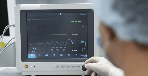
You can record an ECG using electrodes placed on specific body locations. These electrodes detect the impulses generated by the heart and transmit them to a machine, which produces graphical representations of the heart’s electrical activity.
ECG Waves Explained
Understanding the significance of these components is crucial for accurate ECG interpretation.Take a look at the different types of waves you can see in an ECG.
- P-wave: Represents atrial depolarization, or the contraction of the atria.
- QRS complex: Reflects ventricular depolarization, or the contraction of the ventricles. QRS in ECG gives you an insight into the movement of blood.
- T-wave: Indicates ventricular repolarization, or the relaxation of the ventricles.
- PR interval: Measures the time between atrial depolarization and ventricular depolarization.
- QT interval: Measures the duration of depolarization and repolarization of the ventricles.
Interpreting ECG Patterns
ECG patterns can reveal necessary information about the heart’s condition. You can even learn about this in a recognized BLS certification course. Here are some common ECG patterns and their corresponding meanings:
- Normal Sinus Rhythm: The ideal ECG pattern indicates an average heart rate and rhythm.
- Atrial Fibrillation: Characterized by irregular, rapid atrial contractions, this pattern suggests an abnormal heart rhythm.
- Myocardial Infarction: ST-segment elevation or depression, accompanied by Q waves, indicates damage to the heart muscle caused by a heart attack.
- Bundle Branch Block: This pattern shows abnormalities in the heart’s electrical conduction system, affecting the ventricles’ contraction.
Recognizing these patterns can help healthcare professionals diagnose and treat various cardiac conditions effectively.
Clinical Applications of ECG Analysis
ECG analysis has numerous clinical applications and works in various medical settings. Some critical applications include:
- Diagnosing Heart Diseases: ECG is essential in diagnosing arrhythmias, coronary artery disease, and heart attacks. This skill is crucial in critical care and situations that need ACLS skills.
- Monitoring Cardiac Function: ECG monitoring can assess the effectiveness of medications or interventions in patients with heart conditions.
- Preoperative Assessment: ECG analysis is performed before surgery to evaluate a patient’s cardiovascular health and identify potential risks.
- Fitness and Sports Medicine: ECGs help assess athletes and individuals engaged in physical activities to monitor their heart health and detect any underlying cardiac issues.
Read More: Importance of ACLS Course
FAQ’s
- What is the ventricular contraction on ECG?
In an electrocardiogram (ECG or EKG), ventricular contraction is the electrical activity associated with the contraction (systole) of the ventricles, which are the heart’s lower chambers. The ECG graphically represents the electrical events in the heart over time.
- What is the sequence method ECG?
The sequence method in ECG, also known as the standard lead placement, involves attaching electrodes to specific locations on the body to record electrical signals from the heart. The typical sequence is right arm (RA), right leg (RL), left leg (LL), and left arm (LA). This method provides a standardized approach for obtaining ECG tracings, facilitating accurate interpretation and diagnosis of cardiac conditions.
- What is the third wave/complex of the ECG called?
The third wave or complex of the ECG is the “QRS complex.” It represents the depolarization (contraction) of the ventricles. The QRS complex typically includes three waves, Q, R, and S, reflecting the electrical activity as the heart’s lower chambers pump blood. Analyzing the QRS complex is crucial for diagnosing various cardiac conditions and providing valuable insights into the health and functioning of the heart’s ventricles.
- What does the ECG wave tracing represent?
The ECG wave tracing represents the electrical activity of the heart over time. It consists of several waves and intervals, each reflecting a specific cardiac cycle phase. The P wave signifies atrial depolarization, the QRS complex represents ventricular depolarization and contraction, and the T wave indicates ventricular repolarization. Analyzing these waves provides valuable information about the heart’s rhythm, conduction, and potential abnormalities, aiding in diagnosing various cardiac conditions.
- What is the QRS wave in ECG?
The QRS wave in an ECG represents the depolarization and contraction of the heart’s ventricles. It has three distinct components: Q, R, and S waves. Analyzing the QRS complex provides insights into the health and functioning of the ventricles, aiding in diagnosing cardiac conditions.
Read More: Emergency Medical Situations
Conclusion
Understanding the basic principles of ECG waves assists healthcare professionals monitoring their heart health. By recognizing the different components of an ECG and interpreting patterns accurately, medical professionals can diagnose, monitor, and treat various cardiac conditions effectively. ECG waves explained in this article can help you with valuable insights into the heart’s electrical activity, contributing to improved patient care and overall cardiovascular health.