Table of Contents:
- Introduction
- Understanding 12 Lead ECG
- How Does the 12 Lead ECG Work?
- Importance of Proper 12 Lead ECG Placement
- A Complete Guide on 12 Lead ECG Placement
- 12 Lead ECG Placement in Specific Situations
- Capture Accurate Cardiac Data with 12 Lead ECG
Introduction
While there is rising awareness of CPR classes to handle such cardiac crises, opting for a few diagnostic tests will prevent their occurrence. A 12-lead ECG, is one such important test, which allows your healthcare professional to analyze the electrical activity of your heart. Every year, about 805,000 people in the United States experience heart attacks. About 1 in 5 of these events are silent, where the damage is done, but the person is unaware of it.
Given the figure, it is important to know about the proper 12-lead ECG placement to capture the results accurately. In this blog, we will walk you through a detailed look at the placement of each lead, ensuring precise and reliable results. Let’s dive in!
Master ACLS Now
Get ACLS certified with confidence
Understanding 12 Lead ECG
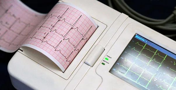
Before delving into the proper 12-lead ECG placement, it’s important to know what the device entails. A 12-lead ECG test is a traditional ECG test that gauges your heart’s electrical activity through electrodes, leads, an amplifier, and a recording device. The test plays a vital role in your heart health, providing insights into heart abnormalities.
Each lead records the heart’s electrical impulses from a specific direction, helping diagnose various cardiac conditions. In the 12-lead ECG, abnormalities are diagnosed through waveform analysis. Therefore, it demonstrates P-wave abnormalities, QRS complex abnormalities, and T-wave abnormalities in the report. If you find any of these abnormalities, the patient might be suffering from a variety of heart conditions, from health palpitations to cardiac arrhythmias and heart attacks.
Read More: What is the difference between Cardiac Arrest and Heart Attack?
How Does the 12 Lead ECG Work?
Understanding the difference between ECG electrodes and ECG leads is important to know the mechanism behind the diagnostic test. Here’s how they are distinguished:
- An ECG electrode is a conductive pad attached to the skin to record the electrical activity of your heart.
- An ECG lead is a graphical representation of your heart’s electrical activity, calculated by analyzing the data from different ECG electrodes.
Remember, a 12-lead ECG records 12 leads that produce 12 separate graphs on a single piece of ECG paper. However, only 10 physical electrodes will be attached to the patient to produce the 12 leads.
Master ACLS Now
Get ACLS certified with confidence
Importance of Proper 12 Lead ECG Placement
Accurate 12 ECG lead placement is vital for diagnosing conditions like myocardial infarction and different classifications of arrhythmia. Even slight deviations can interfere with interpretations, leading to misdiagnosis or missed diagnosis, which affects overall patient care. Proper placement ensures that the electrical activity reflects your true heartbeat and functions. The 12 leads should be strategically positioned to monitor the ventricular and atrial activity, aligned with the complete cardiac performance.
A Complete Guide on 12 Lead ECG Placement
The concept of 12-lead ECG placement might sound intricate, but it’s important to learn the technique if you work in cardiac care. This guide provides step-by-step instructions for placing all ECG components in the test.
Anatomical Landmarks of 12 Lead ECG Placement
For the proper 12 lead placement ECG electrodes, you should learn the anatomical landmarks of the human body. These landmarks indicate the reference points for precise ECG positions. For electrode placement, the fourth intercostal space and the midclavicular line are commonly used. Finding the sternal notch and xiphoid process will ensure the smooth placement of electrodes. It is vital to understand the anatomy of the chest to place the electrodes in the proper positions and get accurate readings in the ECG test.
12 Lead Placement Techniques
The 12-lead ECG system consists of several leads, each showcasing a unique perspective of the heart’s electrical activity. To detect heart abnormalities accurately and capture particular cardiac relations, you should ensure the position of the ECG is accurate. The techniques of lead placement may differ by the lead system being used. You should familiarize yourself with the electrode placement for each lead to get accurate and comprehensive ECG data.
For instance:
- Standard limb leads (I, II, III), which are placed on your limbs.
- Augmented limb leads (aVR, aVL, aVF) which are calculated as a combination of other precordial leads.
- Precordial leads (V1 – V6), which are placed on your torso.
Lead Placement
The limb leads are positioned on the arms and legs, including Leads I, II, III, aVR, aVL, and aVF. Here’s a brief overview of the position of ECG leads 12 lead.
| Area | Position |
| RA (Right Arm) | Place the electrode anywhere between the shoulder and wrist, avoiding bony areas. |
| LA (Left Arm) | Place the electrode anywhere between the shoulder and wrist, similar to the right arm. |
| RL (Right Leg) | Place an electrode on the right leg, just above the ankle or lower leg. |
| LL (Left Leg) | Place an electrode on the left leg, mirroring the right leg. |
Precordial (Chest) Leads
The six precordial leads (V1 to V6) provide a detailed view of your heart’s horizontal plane. Here’s an overview that covers the standard position for electrode placement.
| Precordial Leads | Position |
| V1 | Place the electrode in the fourth intercostal space, just to the right of the sternum. |
| V2 | Place the electrode in the fourth intercostal space, just to the left of the sternum. |
| V3 | Place the electrode between V2 and V4. |
| V4 | Place the electrode in the fifth intercostal space at the midclavicular line. |
| V5 | Place the electrode horizontally, even with V4, at the anterior axillary line. |
| V6 | Place the electrode horizontally, even with V4 and V5, at the midaxillary line. |
Master ACLS Now
Get ACLS certified with confidence
12 Lead ECG Placement in Specific Situations
The acquisition of 12 lead ECGs in the field theoretically does not differ from those obtained in emergency departments. However, due to the unique prehospital environment, there are several tips to consider when placing the electrodes. Let us explore the different 12-lead ECG placements in specific situations.
Electrode Placement for Women
Before the procedure, it is important to explain to the patient that the chest leads may need to be placed around and under the left breast. Ensure that you lift the patient’s breast to position the V3, V4, and V5 electrodes properly.
Electrode Placement for Bariatric Patients
Accurately positioning the ECG 12 lead may initially seem more challenging in obese or overweight individuals. Therefore, it is important to focus on locating anatomical landmarks. Palpate more deeply to identify the sternal border and the Angle of Louis for placing leads V1 and V2. V4 is typically located along a straight line below the chest at the fifth intercostal space.
Next, imagine a line extending straight down the left lateral side of the chest. Along this line, the mid-axillary line will mark the location of lead V6. Once you are clear on the ECG where to put leads, place V3 halfway between V2 and V4 and V5 halfway between V4 and V6.
Electrode Placements for Pregnant Patients
Despite the appearance of the abdomen in pregnant patients, the placement of the 12 ECG lead placement remains the same. Keep in mind that left-axis deviation may appear on the ECG in both pregnant and obese patients due to the abnormal position of the heart as the diaphragm is pushed higher into the thoracic cavity.
Electrode Placements for Pediatric Patients
For 12 lead ECG placement in pediatric patients, it is advisable to use smaller electrodes suited to their size. Using adult-sized electrodes may result in overlapping and significantly inaccurate placement. If the patient is under four years old, having a parent close by can help provide reassurance.
Read More: What Does Normal Sinus Rhythm Look Like on ECG?
Tips for Mastering 12 Lead ECG Placement
Mastering 12 Lead ECG placement requires practice, patience, and, most importantly, attention to detail. The following are some tips to help you develop these skills:
- Regular Training Sessions: Hone your skills through regular training sessions, using simulation tools, and seeking feedback from experienced peers.
- Familiarity with Landmarks and Guidelines: Familiarize yourself with the anatomical landmarks in the human body and stay up to date with the latest lead placement guidelines.
- Stay Updated on Advancements: Stay informed about advancements in ECG technology, particularly the latest portable ECG devices.
- Continuous Learning and Skills Refinement: Engage in continuous learning and skills refinement to master 12-lead ECG placement and ensure the accuracy of ECG tests.
Capture Accurate Cardiac Data with 12 Lead ECG
Mastering 12-lead ECG placement is essential for any cardiologist or healthcare professional involved in cardiac care. With proper practice and preparation, obtaining the correct 12-lead ECG placement becomes a more achievable goal. Familiarize yourself with anatomical landmarks and learn the precise placement of the electrodes on the patient’s body.
Ensure that you carefully adhere to protocols as part of your routine training to obtain the highest quality diagnostic tracings. Additionally, with technological advancements, it is important to learn about portable ECG devices to generate accurate recordings. Remember, precise electrode positioning is a fundamental skill necessary for capturing accurate cardiac data and making informed clinical decisions.


