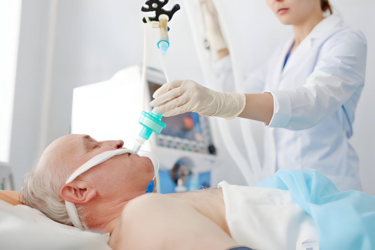
The proper placement of the endotracheal tube secures a patient’s airway during anesthesia administration or resuscitation. Ensuring right ET tube placement prevents complications like hypoxia or trauma. Constructed of polyvinyl chloride, an ET tube should be placed between the vocal cords through the trachea.
There are various techniques used for et tube intubation. It is important for healthcare professionals to learn both primary and confirmatory methods to verify ETT placement. In this blog we will learn about the different monitoring techniques used to place the endotracheal tube.
What are the steps of endotracheal tube placement?
Endotracheal intubation is a vital procedure performed to secure a patient’s airway. This ensures adequate ventilation and oxygenation during surgery and other emergency care. The process requires skill and precision to avoid complications. Here are the essential steps involved in endotracheal tube placement:
- Preparation:
- Gather necessary equipment: Endotracheal tube, laryngoscope, suction device, stylet, bag-valve-mask (BVM), and monitoring devices.
- Pre-oxygenate the patient with 100% oxygen for 3-5 minutes to increase oxygen reserves.
- Position the patient appropriately, typically in the sniffing position, to align the airway axes.
- Pre-Medication (if applicable):
- Administer sedatives and paralytics as needed for comfort and muscle relaxation.
- Laryngoscopy and Visualization:
- Hold the laryngoscope in the left hand and insert it into the patient’s mouth, sweeping the tongue to the left.
- Advance the laryngoscope blade to visualize the vocal cords by lifting the epiglottis.
- Endotracheal Tube Insertion:
- Insert the endotracheal tube through the vocal cords into the trachea, ensuring it is placed just above the carina.
- Remove the stylet (if used) while stabilizing the tube.
- Confirmation of Placement:
- Inflate the cuff of the endotracheal tube to secure it within the trachea.
- Verify placement using multiple methods: auscultate bilateral breath sounds, observe chest rise, and check for the absence of gastric sounds.
- Confirm with capnography by detecting end-tidal CO2.
- Obtain a chest X-ray if necessary for definitive confirmation.
- Securing the Tube:
- Secure the endotracheal tube with adhesive tape or a tube holder to prevent displacement.
- Mark the tube at the level of the teeth or gums to monitor its position.
- Post-Intubation Management:
- Connect the tube to the mechanical ventilator or BVM (Bag Valve Mask).
- Monitor the patient’s vital signs, oxygenation, and ventilatory parameters continuously.
- Adjust ventilator settings as needed based on the patient’s condition and blood gas analysis.
Read More: Study Guide To Body Systems: ACLS Certification Resource
What are the monitoring techniques to ensure proper ET tube placement?
Ensuring the correct placement of an endotracheal tube (ETT) is essential for effective ventilation. Several monitoring techniques are employed to verify the tube’s position within the trachea. This prevents complications like esophageal intubation or accidental extubation. Here are the key monitoring techniques used to confirm proper ETT placement:
- Direct Visualization: Visual confirmation of the tube passing through the vocal cords during laryngoscopy.
- Auscultation: Bilateral auscultation of breath sounds over the lungs to ensure equal and adequate ventilation. Absence of breath sounds over the stomach to rule out esophageal intubation.
- Capnography: Continuous monitoring of end-tidal CO2 (ETCO2) levels, which provides real-time confirmation of tube placement in the trachea. Presence of a consistent ETCO2 waveform indicates correct placement.
- Chest Rise and Fall: Observation of symmetrical chest movement with each ventilation to verify proper lung inflation.
- Tube Markings: Checking the depth markings on the ETT at the level of the teeth or gums to ensure it is at the correct depth.
- Esophageal Detection Devices: Use of devices like the esophageal detector device (EDD) to distinguish between tracheal and esophageal placement.
- Ultrasound: Bedside ultrasound to visualize the tracheal tube placement and confirm bilateral lung sliding.
- Condensation in the Tube: Observation of condensation within the ETT during exhalation, indicating airflow through the trachea.
- Fiberoptic Bronchoscopy: Direct visualization of the carina and main bronchi using a fiberoptic scope to ensure correct placement.
- Chest X-ray: Post-intubation chest X-ray to confirm the tip of the ETT is positioned 2-3 cm above the carina and to check for lung complications.
Read More: Cardiac disease in the young: Recognizing and Understanding Early Signs
Conclusion
Confirming the placement of the endotracheal tube ensures effective ventilation. Monitoring techniques like visualization, chest x ray and auscultation, verifies the placement of Et tube. These monitoring techniques help prevent fatal conditions like hypoxia or pneumothorax. When healthcare professionals master these techniques they ensure optimal patient care along with positive clinical outcomes.